Protocol: Isolation from infected tissue
1. If a lesion is not sporulating or has few sporangia:
A. Place leaflets in a moist chamber (3 or 4 leaflets/plate). Do not crowd the leaves, and make several moist chambers per sample. Keep any unused material in a plastic bag in the refrigerator at 15oC.
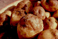
B. Surface sterilize a tuber in 10% Chlorox bleach (for about 5 minutes for a small tuber), and cut it into slices that are 1/8-1/4" thick. Cover a lesion with a potato slice in a petri plate. If the potato slice is thin enough, use a water agar plate—don’t let the tuber slice touch the agar. For thicker slices, use an empty plate.
C. Check plates every 2-3 days for sporulation. When lots of sporangia are available, continue with step 2, substituting the mycelium growing through the tuber for a lesion.
2. If a lesion is sporulating heavily:
A. Brush a small piece of clean-up agar across the sporangia and place on a clean-up agar plate. Up to 4 such pieces of agar may be placed on one plate. Make several such plates per sample.
B. Cut out a section of the lesion (ideally 1 cm diameter) and place in 1.5 ml microfuge tube with 1 ml dH20. Use this for cellulose acetate. Cellulose acetate may also be performed later.
3. After the pathogen is isolated on clean-up agar, it may be transferred to rye A or rye B plates.
4. Transfer 3 plugs of mycelium from rye A or rye B plates to each of two pea broth plates. Once the mycelium has grown up enough, vacuum filter the mycelium, place it at –80oC for at least 2 hours, and lyophilize it overnight. This mycelium will be saved at –20oC for DNA extraction.
5. Transfer the sample to a rye A plate. Set this plate aside, as it will be used for putting the sample into storage.